- MODULE 1: Introduction to ABI
- Module Introduction
- Take the Pre-test
- a) A person’s abilities & life span
- b) What is ABI?
- c) Causes of ABI
- d) Incidence
- e) The brain
- f) Severity of ABI
- g) Cause to impact
- h) Rehabilitation
- i) Rehabilitation stages & pathways
- j) Common effects
- k) Impacts on life
- l) People with ABI
- m) Family and friends
- n) Key messages and
Tools to explore in Module 2 - o) Building skills
- Take the Post-test
e) Understanding the brain
- i) Intro :
Graphic - ii) Skull
- iii) The
Brain Q - iv) Brain
animations - v) Nervous
system Q - vi) Neurons
Q - vii) How it
all works
i) Introduction
Different areas of the brain control different functions.
SLIDES:
To pause: Hover mouse over slide. To continue: move mouse off slide.
To go to a specific slide: Click on slide numbers below.


(c) Copyright - See: Module 1: An Introduction to Traumatic Brain Injury www.TBIStaffTraining.info
ii) The Skull
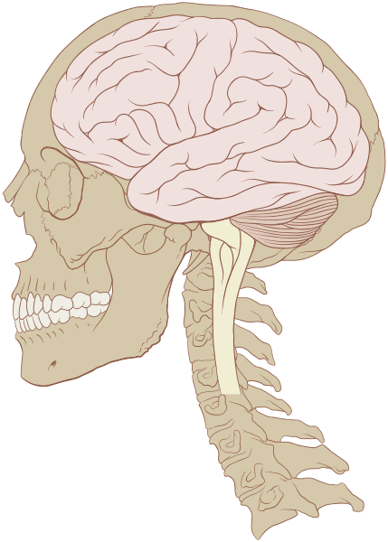
The brain sits within the skull and connects to the spinal cord.
The skull has bony ridges on its inner surface. In the picture below you are looking into the skull with the top of the skull removed.
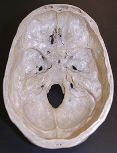
These ridges can cause damage to brain tissue in the event of a TBI.
iii) The brain
The brain is the body's control centre. It may only weigh about 1.5kg but it is made up of billions of cells. It controls everything we do from basic bodily functions such as breathing, heart beat and blood pressure, to our movements, speech, senses and our personality.
Cerebral Hemispheres
The brain is divided into two cerebral hemispheres – the left hemisphere and the right hemisphere. These hemispheres are largely symmetrical in size and structure. The two hemispheres work seamlessly together, processing sensory information from the environment and initiating motor responses.
Generally speaking, the movement on the right side of our body (the actions of our arms and legs) is controlled by the left side of the brain (left hemisphere) and vice versa, movement on the left side of the body is controlled by the right side of the brain (right hemisphere).
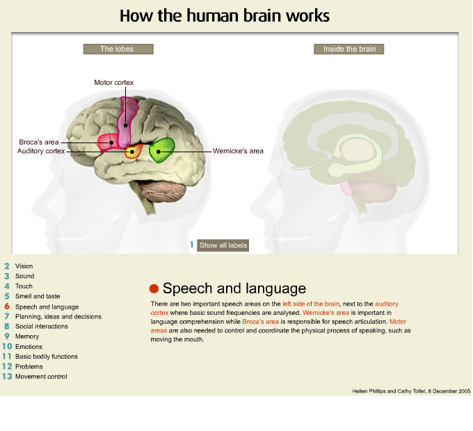
Each hemisphere also has specialised functions. The left hemisphere tends to play a significant role in speech and language. The right hemisphere plays a special role in the perception and integration of nonverbal information including facial recognition.
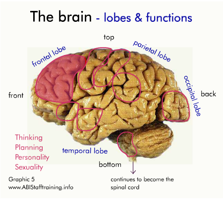
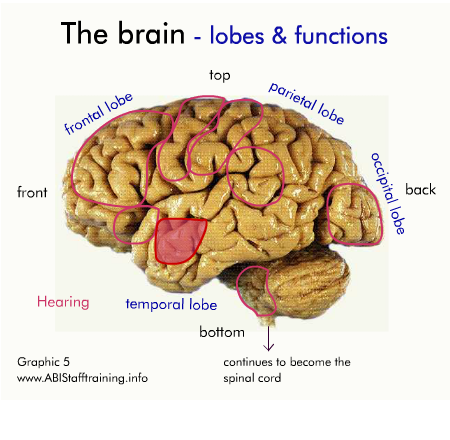
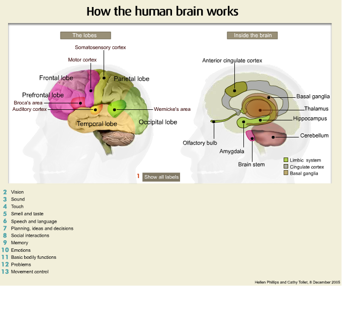
Lobes
The cortex of each hemisphere comprises four ‘lobes’ that handle specific areas of function. The animation immediately below shows the left and right hemispheres and the four lobes (red, green yellow and light yellow) within each hemisphere. (The blue area is the cerebellum.)
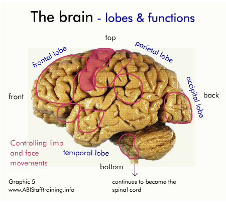
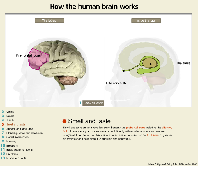
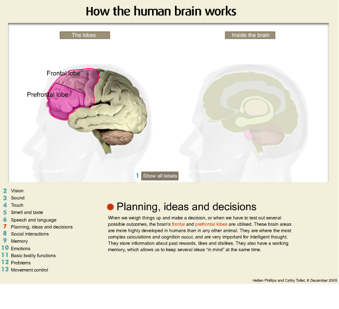
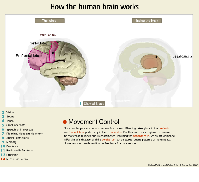
Frontal lobes
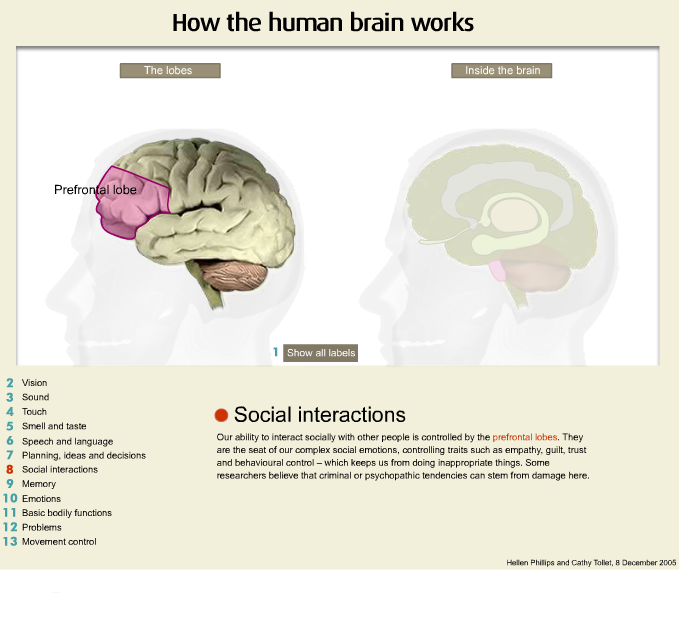
The frontal lobes have different subdivisions. The back division of the frontal lobes are part of the brain’s systems for controlling our movement. The front division of the frontal lobes play an important role in higher-level thinking skills (such as planning, organising, reasoning, decision-making and judgement.), as well as aspects of behaviour, emotions and personality. They rely on complex connections and information exchanges with other areas of the brain to perform these ‘executive functions’. The animation immediately below shows the left frontal lobe.
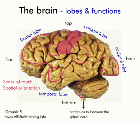
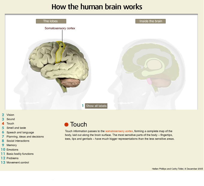
Parietal lobes
The parietal lobes are divided into two zones. The front half of the parietal lobes are principally concerned with receiving data from the bodies’ senses such as touch, pressure, temperature, and pain and then communicating this information to the frontal lobes. The rear section of the parietal lobes deals with spatial awareness, such as our ability to find our way around, and to reach for objects. Interestingly, this region also plays an important role in our ability to do arithmetic.
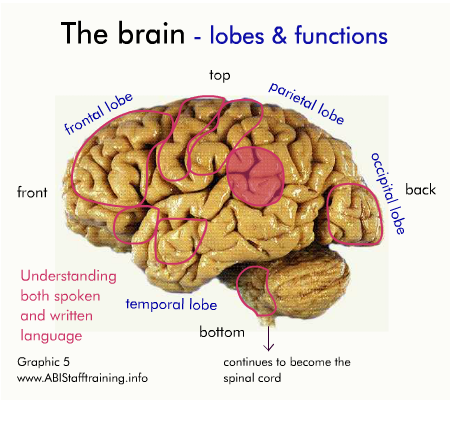
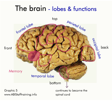
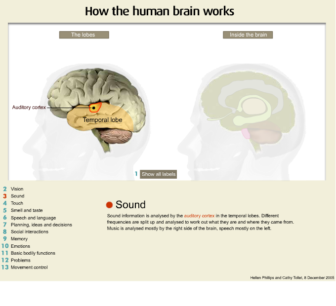
Temporal lobes
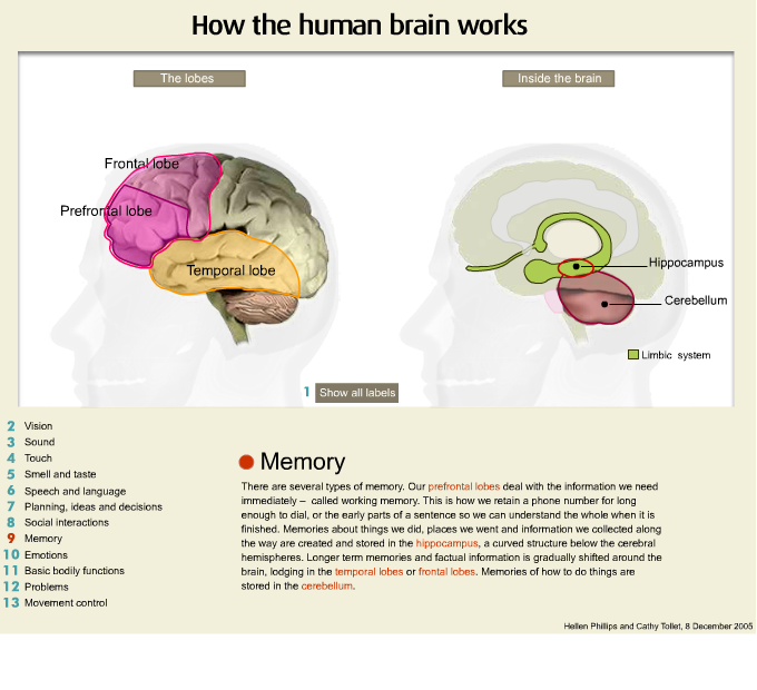
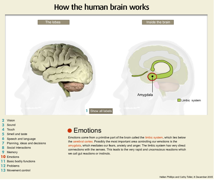
The temporal lobes also have a role in language, particularly in the ability to hear and understand. The temporal lobes are also concerned with memory, the emotions, the ability to enjoy music and to recognise and identify things we see, such as faces or objects.
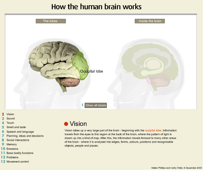
Occipital lobes
The occipital lobes are primarily concerned with vision but also with our ability to recognise what we see in terms of identifying colours, locating objects in the environment and seeing objects accurately.
The animation immediately below shows the left occipital lobe.
Cerebellum and Brain Stem
Below the cerebral hemispheres are the cerebellum and the brain stem, which connect with the spinal cord.
Cerebellum
The cerebellum ‘carries out’ orders from the cerebral hemispheres and keeps a number of vital but routine functions kicking over, such as maintaining balance and ensuring our muscles move in a smooth, coordinated way.
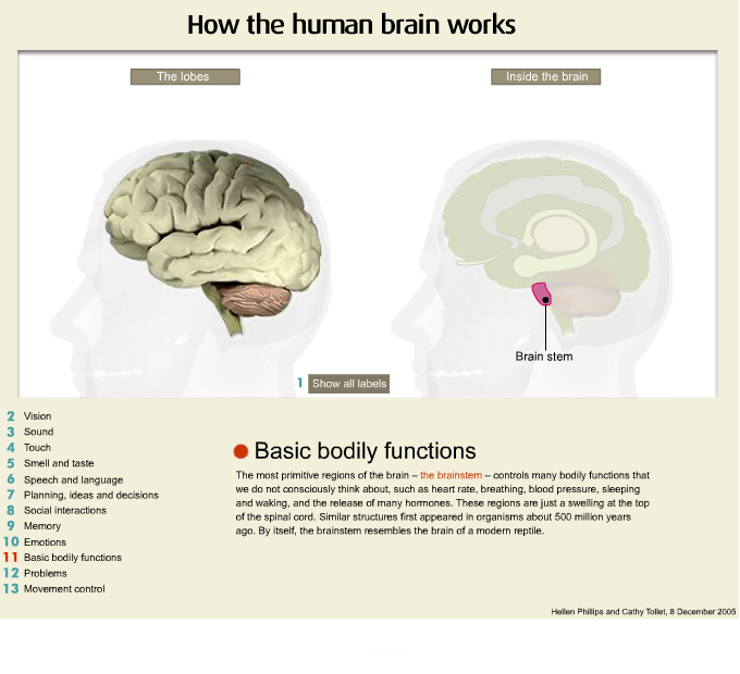
Brain stem
The brain stem controls many vital functions including breathing, blood pressure, blood circulation, swallowing, appetite, body temperature and digestion, as well as the need for water, staying awake and sleeping, among other things. It is also the main route for nerve fibres running between the cerebral hemispheres and the spinal cord. Even small injuries to the brain stem can be life threatening.
Based on Brainlink Fact Sheet 1 Understanding the Brain www.brainlink.org.au and material from the Brain Injury Rehabilitation Directorate
Answer the questions
What a primary function of the parietal lobe?
What 's a primary function of the occipital lobe?
What's a primary function of the frontal lobe?
What a primary function of the parietal lobe?
What 's a primary function of the occipital lobe?
iv Brain animations - How the brain works
SLIDES:
To pause: Hover mouse over slide. To continue: move mouse off slide.
To go to a specific slide: Click on slide numbers below.
v) The nervous system
The brain communicates messages through a complex network of nerves that travels throughout our body. The brain nerves together make up the ‘nervous system’, whilst the brain and spinal cord together constitute the ‘central nervous system’.
On their own, the nerves that run throughout our body are called the ‘peripheral nervous system’. They relay information from the brain through the spinal cord to the rest of the body and back again.
The ‘autonomic nervous system’ is part of the peripheral nervous system. It conveys messages from all of the organs in our chest, abdomen and pelvis. For example, it manages our "fight and flight" responses, and our "rest and digest" responses. It looks after the automatic activities of our heart and blood vessels and plays an important part in sexual response and bladder control.
Based on Brainlink Fact Sheet 1 Understanding the Brain www.brainlink.org.au and material from the Brain Injury Rehabilitation Directorate
Answer the question:
If a person is unable to move their arm - what are three possible causes?
vi) Neurons
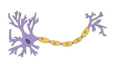
These are the basic components of the nervous system, also called are nerve cells or neurons. Neurons are a special type of cell that process and transmit information by electrical and chemical signaling.
We are born with about 100 billion neurons that must last a lifetime. Unlike all the other cells in the body, neurons usually cannot replace themselves if they die or are damaged.
The ‘grey matter’ of the brain is made up of neuronal cells. It can be found on the surface of the cerebral hemispheres and the cerebellum, as well as in the depths of the cerebrum, cerebellar nuclei, brain stem and inner part of the spinal cord.
‘White matter’ is found in the inner part of the brain and outer part of the spinal cord. It is made up of bundles of myelinated nerve fibres called axons, which connect various grey matter areas to each other, and carry nerve impulses between neurons. These axons are covered by a white, fatty substance called myelin (hence the term ‘white matter’), which insulates them, like the plastic coating of an electric wire. Myelin acts as an insulator, increasing the speed of transmission of all nerve signals. The axons then bundle together, like the individual telegraph wires in a cable, to form a nerve.
Based on Brainlink Fact Sheet 1 Understanding the Brain www.brainlink.org.au and material from the Brain Injury Rehabilitation Directorate
Answer the question
When neurons die are they replaced?
vii) How it all Works
The brain is in constant contact with all parts of the body, sending instructions and receiving feedback from the senses. The axons carry these messages as tiny electrical currents or nerve impulses.
Outgoing messages
Messages sent from the brain to activate the muscles of the body travel along the motor pathways. The neurons that make up these pathways are called motor neurons.
Incoming messages
Incoming messages: Messages sent from the senses back to the spinal cord and brain come along the sensory pathways. These are called sensory neurones.
Damage to the brain may affect many different functions and abilities. An injury can result in a serious disability that will interfere with a person’s daily functioning and personal activities, often for the rest of their life. While the outcome of the injury depends largely on the nature and severity of the injury itself, long-term effects are difficult to predict and will be different for each person. It is common for people with a brain injury to get tired more quickly, have difficulty with short-term memory and find it more difficult to concentrate and to remember information.
The areas in which people with ABI may experience long-term changes include:
- Medical difficulties (e.g., epilepsy)
- Changes in physical and sensory abilities (e.g., weakness, tremor, impaired vision or smell)
- Changes in the ability to think and learn (cognition)
- Changes in behaviour and personality (psychological)
- Communication difficulties (e.g., slow or slurred speech , difficulty following conversation).
Based on Brainlink Fact Sheet 1 Understanding the Brain www.brainlink.org.au and material from the Brain Injury Rehabilitation Directorate